TEL:0086-20-34174605/34174486
MOB:0086-18998432898(WhatsApp available)
E-mail:[email protected]
- Home
-
Products
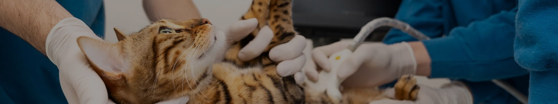
Veterinary LaboratoryHematology Analyzer Chemistry Analyzer Coagulation Analyzer Urine Analyzer Electrolyte Analyzer Microplate Reader & Washer Microscope Centrifuge Animal Sperm Quality Analyzer Veterinary Roller Mixer / Shaker Vet Clinic Water Bath Vacuum Blood Collection Tube Veterinary Pipette Immunofluorescence Analyzer Blood Gas and Chemistry Analyzer Veterinary Progesterone Analyzer Veterinary Sperm Analyzer
- Download
- Reviews & References
- News
- About Us
- Contact Us
- Blog